A number of active projects on Folding@home right now aim to understand how different forms of the protein apolipoprotein E (ApoE) determine one’s risk of developing Alzheimer’s disease.
Alzheimer’s disease is the 6th leading cause of death in the USA and there are no effective treatments. Moreover, the prevalence of this age-related neurodegenerative disease is likely to increase as the population ages. Therefore, there is a great need to understand Alzheimer’s disease and develop therapeutics.
ApoE is an appealing target for treating Alzheimer’s disease because which form of this lipid transporter a person has is one of the best predictors of how likely they are to develop Alzheimer’s. People with the ApoE4 form are up to 15-fold more likely to develop Alzheimer’s disease than people with the more common ApoE3 form. Meanwhile, people with ApoE2 appear to have a lower risk of developing Alzheimer’s. However, the mechanism coupling ApoE and Alzheimer’s disease remains unclear.
Understanding the structural differences between the different forms of ApoE could enable the design of ‘structure correctors’ that combat Alzheimer’s disease by stabilizing non-toxic conformations. However, characterizing these differences remains challenging. ApoE is extremely dynamic, so it can’t be studied with most experimental techniques for determining a protein’s structure.
That’s where Folding@home comes in. We are currently simulating with various forms of ApoE to understand what makes them different.
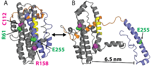
Our results on ApoE4 are quite surprising. It has long been believed that the two ends of ApoE4 interact with each other in a way that ApoE3 and ApoE2 do not. This unique interaction in ApoE4 has been proposed to somehow trigger other processes in the brain that lead to Alzheimer’s disease. However, we don’t see any evidence for this interaction! Moreover, experiments on single ApoE proteins are consistent with our computational predictions. Hopefully, the simulations that are currently running on Folding@home will shed light on what’s really going on.